Services at Aurora Biolabs
Assay Development & Assaying Services
Scientists at Aurora Biolabs (an SBD company) and Structure Base Design, Inc. (SBD), have been providing assay development and assaying services to pharmaceutical/biotech companies and academia institutions for almost two decades. Our clients include Quidel, Mirati Therapeutics, Tanabe Research Laboratories, Caltech, and Salk Institute. We have consistently delivered quality results for our clients and partners. Many of our customers have submitted compounds discovered at Aurora Biolabs and SBD for clinical trials. The assays listed below, by no means exhaustive, are the assays that have been well-established at Aurora Biolabs. We also customize assays and develop de novo assays tailoring to each customer’s project and technical requirements.
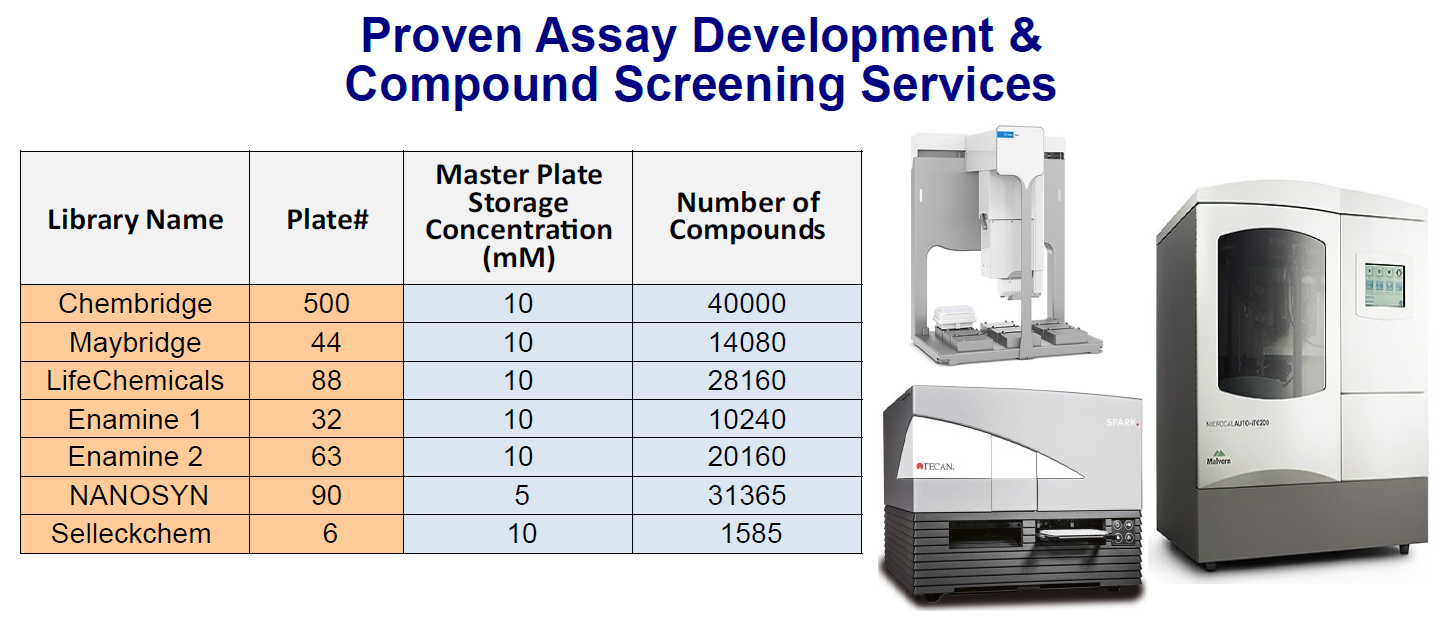
HTP Compound Library Screening With 145K Compounds
Structure-Based Drug Discovery (SBDD) is a well-established method for drug discovery. With the availability of increasingly powerful computers and ever-growing big data in the drug discovery space, SBDD is arguably the most efficient and powerful drug discovery process. Fragment-Based Drug Discovery (FBDD) evolved from the de novo drug design approach to further speed up the drug discovery process. Since the first Fragment-Based Drug Discovery (FBDD) drug, Zelboraf® (vemurafenib, PLX4032) was approved by FDA in 2011, there are now over 6 FDA-approved FBDD drugs on the market and 40+ FBDD drug candidates in clinical trials.
At Aurora Biolabs, an SBD company, we provide services in both SB-CADD (Structure Based-Computer Assisted Drug Discovery) screening and FBDD screening on more than 145,000 compounds. SBD can also provide crystallization service for drug target proteins and co-crystallization of drug target-compound. Contact Us for detail.
Kras Ligand Exchange Assay
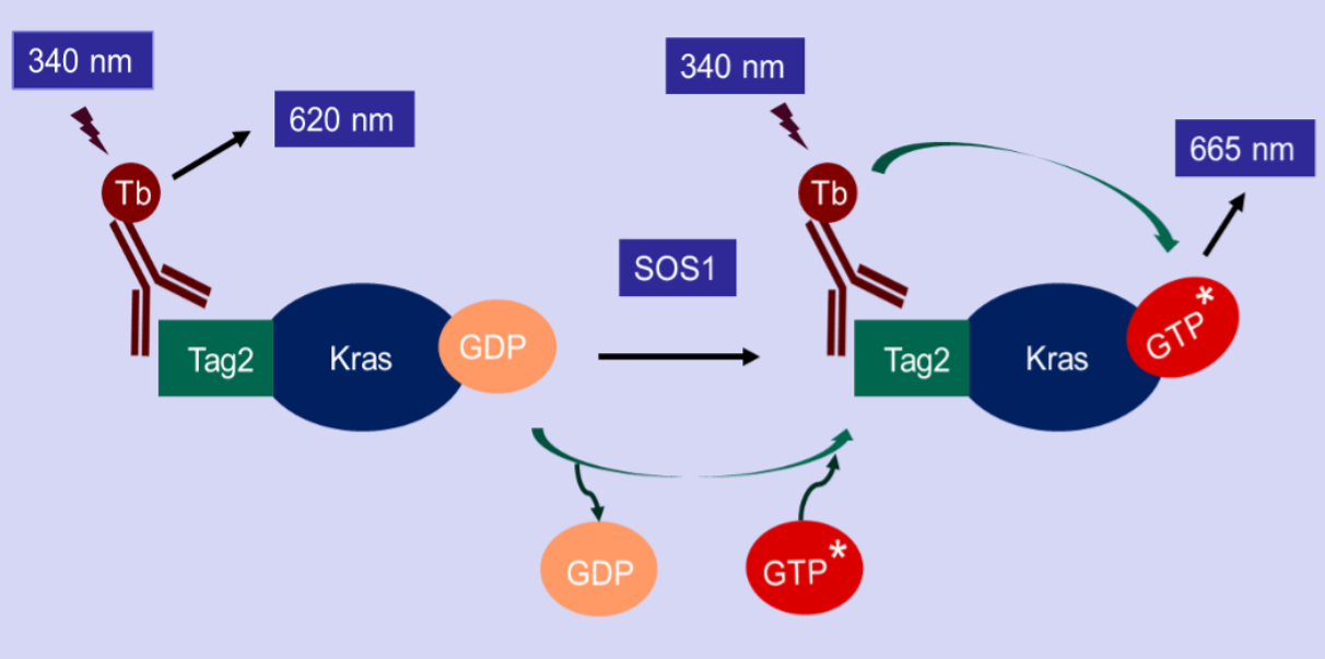
Aurora Biolabs’ nucleotide exchange assay is a TR-FRET based assay. The assay kit is designed to detect the GTP binding status of Kras in the presence of SOS1, the most-studied guanine nucleotide exchange factor (GEF) of Kras. The Tag2-Kras in this assay kit is recognized by a Terbium-labeled anti-Tag2 antibody (HTRF donor). If Kras binds to a fluorescence-labeled GTP (HTRF acceptor), the donor and the acceptor will be brought in close proximity. Excitation of Terbium (340nm) generates fluorescence resonance energy transfer (FRET) to the fluorescence-labeled GTP acceptor, which consequently fluoresces at 665 nm (figure below). Thus, GTP binding to Kras can be quantitively measured by calculation of the fluorescent ratio of 665 nm/620 nm. The inhibitor blocking the nucleotide exchange will reduce the HTRF signal.
Kras-cRAF Binding Assay
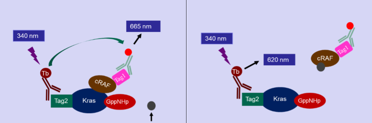
The Kras-cRAF binding assay is a TR-FRET based assay. In this assay, Kras is loaded with GppNHp,representing the activated Kras. The assay kit is designed to detect binding between Kras and cRAF.
The Kras in this assay kit has a Tag2, that can bind to a Terbium-labeled anti-Tag2 antibody (HTRF donor), and cRAF in this assay kit has a Tag1, that can bind to a fluorescence-labeled anti-Tag1 antibody (HTRF acceptor). The binding of Kras with cRAF results in fluorescence resonance energy transfer (FRET) from the HTRF donor to the HTRF acceptor when the donor is activated allowing cRAF binding to be measured.
PKMYT1 Binding Assay
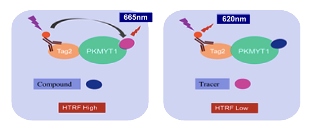
PKMYT1 is a membrane-associated tyrosine- and threonine-specific cdc2 inhibitory kinase. It belongs to WEE1 family of kinases that play an important role in the regulation of mitosis. Aurora Biolabs’ PKMYT1 Binding Assay kit is a TR-FRET based assay designed to screen compounds that bind to PKMYT1. Binding of a fluorescence-labeled tracer by PKMYT1 results in fluorescence resonance energy transfer (FRET) between the fluorescence donor bound to PKMYT1 and the fluorescence acceptor on the tracer, resulting in a fluorescence emission at 665nm. Competitive binding of a compound will displace the tracer, thereby resulting in no resonance energy transfer and reducing the receptor signal.
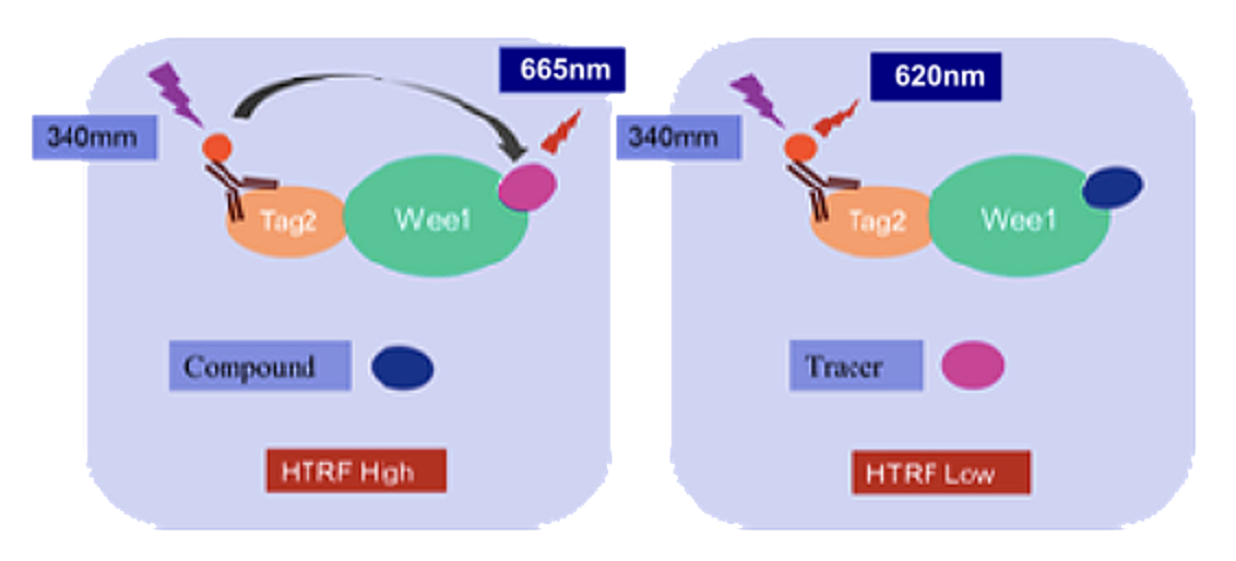
WEE1 Binding Assay
WEE1 is a nuclear kinase that belongs to the WEE kinase family. It negatively regulates the cell cycle via phosphorylation of CDK1 and ahs been identified as a novel drug target to treat cancer. Aurora Biolabs’ WEE1 Binding Assay is a TR-FRET based assay designed to screen compounds that bind to WEE1. Binding of a fluorescence-labeled tracer by WEE1 results in fluorescence resonance energy transfer (FRET) between the fluorescence donor bound to WEE1 and the fluorescence acceptor on the tracer, resulting in a fluorescence emission at 665nm. Competitive binding of a compound to WEE1 results in no fluorescence energy transfer, and thus reducing the receptor fluorescence signal.
TR-FRET Custom Assay Development Services
A Time-Resolved Förster Resonance Energy Transfer (TR-FRET) assay is a biochemical technique that combines the principles of Time-Resolved Fluorescence (TRF) and Förster Resonance Energy Transfer (FRET) to achieve high sensitivity and reduced background noise. It is a powerful tool to study molecular interactions — such as protein–protein, receptor–ligand, or protein–nucleic acid binding — in a homogeneous (no-wash) format.
Principle of TR-FRET
FRET is a phenomenon where energy is transferred from a donor fluorophore to an acceptor fluorophore when they are within very close proximity (typically less than 10 nanometers).
Two lanthanide complexes, europium and terbium, are commonly used donor fluorophores. When the donor is excited by light (LED or Laser) at 325 nm or 340 nm, it can transfer energy to the acceptor if they are within very close proximity (typically less than 10 nanometers), leading to acceptor fluorescence at a different wavelength (emission wavelength depending on the acceptor fluorophore used).

Advantages of TR-FRET Assay
Traditional FRET assays can suffer from high background fluorescence caused by interfering compounds and the short-lived emission from standard fluorophores. In the time-resolved florescence (TR-FRET) method, fluorescence is measured after a short delay (typically microseconds) following excitation, since lanthanides have a fluorescence lifetime that is much longer (microseconds to milliseconds) than the nanosecond-range fluorescence of background molecules. TR-FRET eliminates background fluorescence from biological samples and improves the signal-to-noise ratio.
TR-FRET assays are homogeneous high through-put assays with "mix-and-read" format, and do not require a physical separation or wash step to remove unbound labeled molecules, making them simple and efficient for automation.
ADP-Glo Assay
ADP-Glo assay is a Promega assay that measures ATP converted from ADP from a kinase reaction. The increase of luminescence corresponds to the increase of kinase activity. We provide services in performing high throughput screening using ADP-Glo assay. Contact Us for detail.
Fluorescence Polarization Assay
Fluorescence Polarization (FP) measure changes in light polarization that occur when a small fluorophore interacts with or dissociates from a much larger partner. It allows rapid and quantitative analysis of a variety of molecular interactions and enzyme activities in a high throughput format. Scientists at Aurora Biolabs and SBD have decades of experience designing and running FP assays in a high throughput format. Contact Us for more detail.
SPR Binding Assay
Surface Plasmon Resonance (SPR) has evolved from measuring protein-protein interaction to measuring affinity between small analyte (<1 kDa) to macromolecule ligand (>10 kDa). It is widely used in fragment-based drug screening in the pharmaceutical industry. At Aurora Biolabs and SBD, we have close to 2 decades of experience serving the pharmaceutical industry in drug discovery programs. Contact Us for more detail on our SPR binding assay services and what we can offer to speed up your drug discovery campaigns.
Cell-Based Assay
Cell proliferation analyses are crucial for cell growth and differentiation studies, and are often used to evaluate both compound toxicity and the inhibition of tumor cell growth during drug development. Proliferation measurements are typically made based on average DNA content or on cellular metabolism parameters.
The Aurora Biolabs’ One Solution Cell Proliferation Assay is a colorimetric method for determining the number of viable cells in proliferation, cytotoxicity, or chemosensitivity assays. The Aurora Biolabs One Solution Reagent contains a tetrazolium compound [3-(4,5-dimethylthiazol-2-yl)-5-(3-carboxymethoxyphenyl)-2-(4-sulfophenyl)-2H-tetrazolium, inner salt; MTS] and an electron coupling reagent (phenazine ethosulfate; PES). PES has enhanced chemical stability, which allows it to be combined with MTS to form a stable solution. The Aurora Biolabs Assay uses phenazine methosulfate (PMS) as the electron coupling reagent, and PMS Solution and MTS Solution are supplied separately. PES has enhanced chemical stability, which allows it to be combined with MTS to form a stable solution.
Assays are performed by adding a small amount of the Aurora Biolabs One Solution Reagent directly into culture wells, incubating for 1–4 hours and then recording absorbance at 490nm with a 96-well plate reader. The quantity of formazan product as measured by the amount of 490nm absorbance is directly proportional to the number of living cells in culture.
Features & Benefits
Simplify Colorimetric Viability Assays: "Add-incubate-measure" format (single-step reagent addition) enables design of homogeneous high-throughput screening assays.
Use a Single Solution: Use as a single solution, filter sterilized and ready to add to assay plates (unlike MTT).
Perform Fewer Steps: Perform the assay in 96-well plates with no washing or cell harvesting. Also eliminates solubilization steps normally required for MTT assays.
Gain Flexibility: Plates can be read and returned to incubator for further color development (unlike MTT).
Avoid Organic Solvents: Requires no volatile organic solvent to solubilize the formazan product (unlike MTT).
Non-Radioactive: Requires no scintillation cocktail or radioactive waste disposal (unlike [3H]-thymidine incorporation assays).
For more information or a quotation, Contact Us for detail.Contact Us for detail.
Protein Expression & Purification
Recombinant Protein production is a powerful technique used in the life sciences. The ability to express and purify a desired recombinant protein in large scale will allow scientists to explore a wide range of possibilities in protein research.
Aurora Biolabs services can take your project directly from gene synthesis to protein expression and recombinant protein purification. Protein products usually are purified with three different columns including affinity, ion exchange, and size-exclusion. The purity of the final products is greater than 95% and can be directly used for crystallization and other high-quality assays.
Specific activities and conformational homology of a protein are also major issues we consider during purification. We not only use general protecting reagents during the purification process, but also add various protection additives based on published literatures about the protein. Protein characterization of the final product can be tested in house or at the local Bioscience Service Center using Electrophoresis, Bioactivity study, Reversed-phase HPLC, Dynamic Light Scattering, Mass Spectroscopy Instruments, and BioCore.
Combining Fermenter, Akta Explorer, and large sized chromatography systems in the laboratory, we can produce quantities of protein samples ranging from 10 mgs to a few grams.
For more information and quotation, Contact Us for detail.
Protein/Antibody Conjugation
Aurora Biolabs also provides protein conjugation services to link your protein and antibody to a variety of substrates, including synthetic polymers, nucleic acids, small molecules, other functional or carrier proteins, latex particles, fluorescent particles, and magnetic particles of various sizes. We are capable of processing bio-reactive conjugates from microgram to gram scale. For more information and quotation, Contact Us for detail.
ADC Development & Optimization
Antibody-Drug Conjugates (ADC) has emerged as the next-generation targeted cancer therapeutics. Its meteoric rise has pharmaceutical companies vying to acquire ADC capabilities through acquisitions or investing heavily to develop ADC programs. The concept of ADC is simple: attach a cytotoxic payload to a monoclonal antibody and release the payload upon binding. However, each component of an ADC: antibody, linker, and payload, requires extensive expertise and experience to develop, optimize and combine into a successful ADC drug. Scientists at Aurora Biolabs and SBD have extensive experience in drug discovery and optimization programs. We have worked on more 100 projects and close to 50% of drug candidates have advanced to clinical trials. Contact Us to learn how we can make your ADC development a success!